A common search for freedom from glasses reveals multiple surgical procedures, each with its prerequisites and recovery timeline. Standard LASIK or PRK procedures can reduce lens dependence, but they may not suit thin corneas, dry eye conditions, or active lifestyles, while implants present their trade-offs. Discover how to evaluate vision correction surgery by assessing corneal thickness, tear film stability, occupational hazards, and daily visual needs, match those with laser or lens techniques, the final choice supports clarity and comfort.
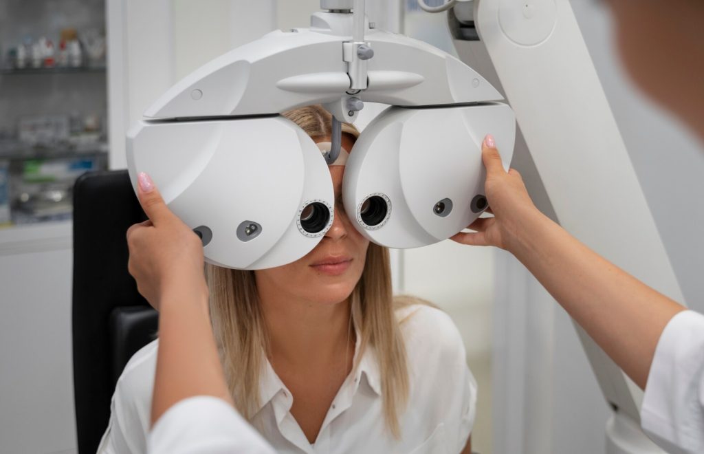
1. The Foundation Exam: Corneal Thickness and Tear Film as Gatekeepers
Before comparing procedure names, the real question is whether the eyes can safely host any vision correction surgery. A thorough pre-operative workup uses advanced diagnostic tools to map the cornea’s shape, measure its thickness, and evaluate tear film stability. These numbers do not just inform the surgeon; they actively disqualify certain options and steer candidates toward safer alternatives.
Corneal thickness and the 500‑micrometre rule: LASIK requires a minimum residual stromal bed of 280 micrometres after flap creation, meaning total corneal thickness typically must exceed 500 micrometres. Candidates below this threshold face ectasia risk. Pentacam tomography reveals central thickness, thinnest point, posterior elevation, and Kmax. Posterior elevation above 25 micrometres or abnormal Kmax flags forme fruste keratoconus, which automatically disqualifies all laser‑based procedures. Surgeons ignoring these values should raise concern.
Tear film quality predicts post‑operative comfort: The OCULUS system measures break‑up time; values under five seconds indicate significant instability requiring at least one month of pre‑treatment with artificial tears and warm compresses. Proceeding without addressing dry eye invites months of grittiness and fluctuating vision.
Interaction between thickness and dryness: Thin corneas combined with borderline dry eyes point away from LASIK toward PRK or ICL. PRK preserves corneal nerves better than LASIK; ICL avoids nerve disruption entirely, making it optimal for both limited thickness and significant dryness.
A proper foundation exam transforms the decision from picking a brand name into understanding which option respects the eye’s inherent limits.
2. LASIK versus PRK: Matching Speed and Safety to Lifestyle
LASIK and PRK both use excimer lasers but take different paths. LASIK creates a flap for faster recovery; PRK removes surface cells, leaving no flap and offering better impact resistance. The choice depends on how soon normal activities must resume and what physical risks daily life presents.
LASIK offers rapid recovery with a permanent flap: Most patients see clearly within 24 hours and return to work next day. Femtosecond lasers create thinner, more predictable flaps than older blades. However, the flap never fully heals to original strength; a strong blow can theoretically dislodge it, though rarely. For fastest visual rebound, LASIK remains dominant.
PRK eliminates flap risks but demands patience: No incision makes PRK suitable for contact sports or jobs with facial trauma risk. The trade‑off: significant burning, tearing, and light sensitivity for the first two to three days. More critically, PRK requires three to four months of topical steroid drops to prevent corneal haze (inflammation‑induced scarring). These steroids can raise intraocular pressure, needing regular monitoring. Missing follow‑ups or stopping drops early risks permanent clouding. Visual clarity improves over weeks, not days.
Lifestyle drives the final recommendation: Patients unable to accept any flap risk choose PRK for peace of mind despite slower recovery. Those needing immediate return to driving and screen work prefer LASIK.
Choosing between LASIK and PRK means weighing immediate convenience against a longer, more medication‑intensive recovery.
3. ICL as a Reversible Anchor: For High Prescriptions and Thin Corneas
Laser‑based correction hits limits above 10 dioptres or when corneal tissue is insufficient. Implantable Collamer Lenses offer an alternative that removes no corneal tissue and preserves natural structure. The lens sits behind the iris, functioning like a permanent contact lens.
Reversibility has practical limits: ICL can be removed or exchanged, but this requires another surgical session with its own risks. The latest EVO ICL has a central hole for fluid circulation, eliminating iridotomy. While reversible in theory, patients should view it as a long‑term implant, not a temporary trial.
Anterior chamber depth is the absolute gatekeeper: ACD must exceed 2.8 millimetres; shallower chambers make implantation anatomically unsafe. Surgeons offering ICL without measuring ACD or accepting borderline values are dangerous. Even with proper ACD, improper lens sizing can cause cataract (touching the natural lens) or angle‑closure glaucoma (excessive vault pressing on drainage structures).
Dry eye outcomes favour ICL significantly: Corneal nerves remain intact, so post‑operative dry eye occurs far less than with LASIK or PRK. For patients with contact lens intolerance or borderline tear tests, this advantage often tilts the recommendation. Contrast sensitivity also remains superior.
High myopia and astigmatism coverage: ICL corrects up to minus 18 dioptres and astigmatism with toric versions. For laser‑ineligible patients, ICL is often the superior choice even when laser remains possible.
4. Wavefront Customisation: Fine‑Tuning for Night Vision and Clarity
Even with the correct procedure, the ablation pattern quality determines whether final vision feels sharp or merely adequate. Wavefront‑guided systems capture unique imperfections beyond sphere and cylinder, reducing night‑time glare and halos.
Distinguishing real customisation from basic treatment: Standard treatments correct routine errors. Wavefront‑guided systems like Contoura use ray‑tracing to reduce starbursts and glare, most noticeable in dim conditions. Basic wavefront‑optimised adjusts spherical aberration but does not fully correct higher‑order irregularities. The difference appears during night driving or high‑contrast viewing.
The cost and tissue trade‑off is real: Wavefront‑guided treatments remove 10‑15% more corneal tissue, critical for borderline thickness patients. Costs run 20‑30% higher. Best candidates have large pupils, frequent night driving, or measurable higher‑order aberrations on diagnostic maps. Those with normal pupils and no night complaints gain little benefit.
Who truly needs customisation: Large pupils allow more peripheral light, making aberrations more noticeable. Wavefront becomes valuable when baseline testing shows significant coma or trefoil. Surgeons should show diagnostic printouts and explain which irregularities the custom treatment addresses. Clinics unable to articulate benefits clearly likely offer minimal customisation with premium branding.
Investing in wavefront‑guided surgery prioritises night performance, but only for those whose maps actually show the need.
Conclusion: Quick Decision Checklist for Vision Correction Surgery
Before committing, review this checklist of key measurements and trade‑offs.
- LASIK: Requires total corneal thickness ≥500 µm and residual stromal bed ≥280 µm after flap. Posterior elevation ≤25 µm with normal Kmax. Tear break‑up time ≥5 seconds preferred. Corrects up to ‑8.00 D. Recovery 1‑2 days; steroid course 1‑2 weeks. Small flap‑related risks exist. No cataract risk. Moderate cost.
- PRK: Requires total thickness ≥450 µm; no residual bed requirement. Same posterior elevation and Kmax criteria as LASIK. Tear break‑up time ≥5 seconds preferred. Corrects up to ‑8.00 D. Recovery involves 5‑7 days discomfort; sharp vision takes weeks. Steroid drops needed for 3‑4 months with pressure monitoring. No flap or cataract risks. Moderate cost.
- ICL: No corneal thickness requirement, but anterior chamber depth must exceed 2.8 mm – non‑negotiable. No posterior elevation criteria. Most forgiving for dry eyes. Corrects up to ‑18.00 D (including astigmatism). Recovery 2‑3 days. No steroids needed. No flap risk, but small cataract risk related to lens sizing. Highest cost.
Use this list during consultation to verify the surgeon’s recommendation matches diagnostic numbers. Any conflict deserves a second opinion. The right procedure fits the eye’s measurements first, preferences second – never the reverse.